Feb. 18, 2026
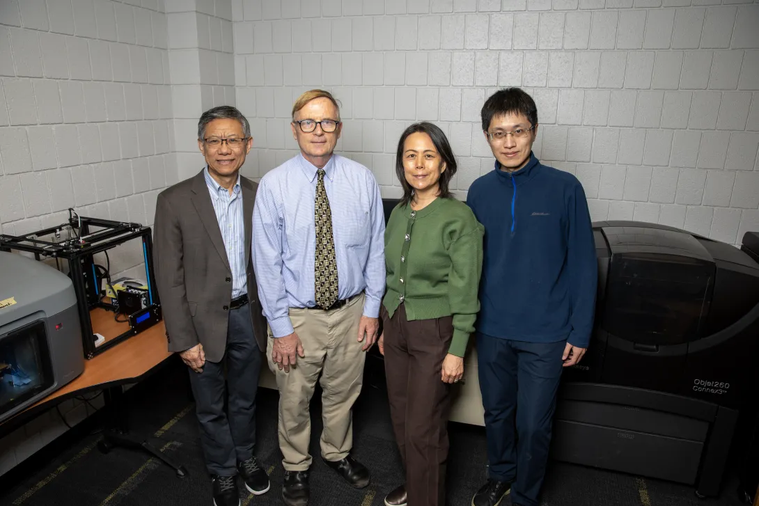
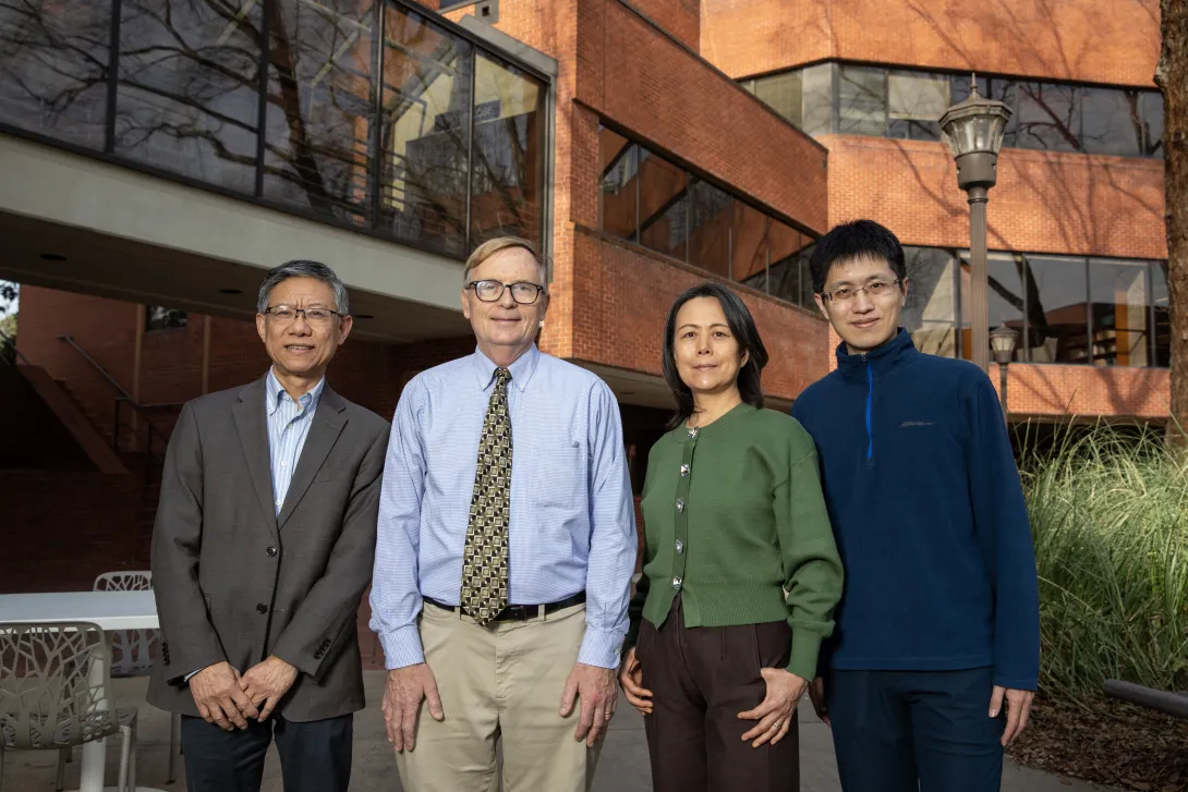
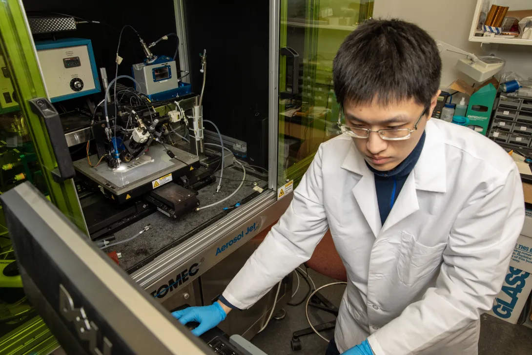
A photo of some of the researchers leading the project written about.

GTRI and Georgia Tech's smart bandage could revolutionize wound care by enabling real-time insights on healing and reducing invasive bandage changes.

A photo of some of the researchers leading the project written about.
A photo of a Georgia Tech graduate student operating the Aerosol Jet® printer to fabricate the sensor.
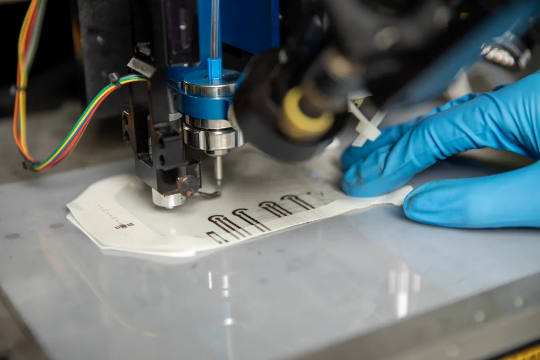
A close-up of the Aerosol Jet printer as it designs a sensor prototype.

While most people don’t think twice about a cut or scrape, for those with diabetes, every wound is a potential threat that requires vigilant care. 

Diabetic foot ulcers, for example, are slow to heal and can increase the risk of infection, hospitalization, and even amputation. 

To address this critical challenge, researchers at the Georgia Institute of Technology (Georgia Tech) and the Georgia Tech Research Institute (GTRI) have developed a sensor designed to monitor chronic wounds in real-time. Embedded directly into a bandage, this flexible, low-cost device could transform wound management for diabetic patients and other critical applications — such as providing direct treatment to soldiers on the battlefield or managing chronic wounds in elderly populations and patients with limited healthcare access — by reducing invasive bandage changes and ensuring timely medical intervention.

“For diabetic patients with foot ulcers, long-term monitoring and care are essential,” said GTRI Principal Research Engineer and Project Lead Judy Song. “We were inspired by the success of wearable glucose monitors to develop a compact, affordable sensor tailored to wound care.”  

This project was supported by GTRI’s Independent Research and Development (IRAD) program between 2022-2025 and reflects the strength of interdisciplinary collaboration across Georgia Tech. Researchers from three out of GTRI’s eight laboratories developed the sensor with experts from the George W. Woodruff School of Mechanical Engineering, the H. Milton Stewart School of Industrial and Systems Engineering and the Wallace H. Coulter Department of Biomedical Engineering at Tech and Emory University.

About one in four people with diabetes will develop a foot ulcer at some point in their lives, making it one of the leading causes of foot amputations. For these patients, nerve damage and poor blood flow hinder the body’s natural healing process and allow wounds to linger and worsen. 

During the initial phases of their research, the team noted that nitric oxide (NO) had been previously identified as a key biomarker for wound health due to its central role in the healing process. Nitric oxide improves blood flow, reduces inflammation, promotes tissue growth and fights infection. By tracking nitric oxide levels in wounds, clinicians could determine whether a wound is improving or detect early signs of trouble. 

"Nitric oxide plays a fascinating, almost paradoxical, role in wound healing,” said GTRI Senior Research Engineer Victoria Razin, who is co-leading the project. “It’s essential for processes like blood flow and tissue repair, but can also signal when something is going wrong.”

At the core of the smart bandage is a flexible sensor powered by a three-electrode system capable of detecting changes in nitric oxide. The team used advanced Aerosol Jet® printing techniques to fabricate the sensor, significantly reducing production costs from thousands of dollars to just a few dollars per unit and making the design more affordable and scalable.

“Typically, prototyping these sensors can cost thousands of dollars, but our approach brought costs down dramatically,” said Chuck Zhang, the Eugene C. Gwaltney, Jr. Chair and Professor in ISYE and a program director at the National Science Foundation (NSF), who oversaw sensor fabrication for this project. “Lower costs let us iterate quickly and deliver something that could have real healthcare impact.”

To test the sensor’s accuracy, the team conducted extensive laboratory studies in both biological and simulated wound conditions. 

In one set of experiments, endothelial cell cultures were used to create “wounds” by scraping the cell layers. As the cells migrated to repair the gap, nitric oxide production increased, and the sensor successfully tracked these changes in real-time. Additional fluid tests using blood plasma and red blood cells demonstrated that the sensor could reliably detect nitric oxide in a variety of conditions that closely mimic real-world wound environments.

These experiments confirmed that the sensor can identify the fluctuations in nitric oxide associated with different phases of wound healing. 

Lab testing was led by Dr. Wilbur Lam, a professor in the Department of Biomedical Engineering and at Emory University School of Medicine, with support from Kirby Fibben, a biomedical engineering Ph.D. student at Tech. 

"There’s a significant clinical need for real time, minimally invasive sensor technologies that detect nitric oxide,” said Dr. Lam. “While we’re starting with wound healing, there’s multiple other applications for vascular, hematologic, and pulmonary diseases as well.” 

The next step in the project is integrating the sensor into a functional wearable device. The team is combining the sensor with a miniaturized potentiostat (MicroPS) – a small electronic device that measures chemical signals – along with flexible electronic components and a system to transmit data to a mobile app. 

The MicroPS, designed by the GTRI research team, led by GTRI Research Engineer Curtis Mulady, enables compact electrochemical measurements and the wireless platform transmits nitric oxide readings from the bandage to a mobile app via Bluetooth. The app uploads the data to a cloud platform, giving clinicians the ability to remotely monitor wound progress in real time. This system could reduce the need for frequent in-person checkups, enabling earlier interventions and improving outcomes for patients.

Future iterations of the bandage aim to include “closed-loop” systems capable of both monitoring and treating wounds, said GTRI’s Song. For example, sensors could trigger a response, like releasing therapeutic agents or antimicrobials directly to the wound, when abnormalities are detected.

The researchers are also exploring commercialization pathways, including partnerships with medical device companies or the formation of a startup. 

“This sensor meets a real need for early detection of infection and to evaluate wound healing, and I believe it could have significant commercial success,” said Peter Hesketh, a professor in the School of Mechanical Engineering who led sensor design and performance testing. 

Other contributors to this project from GTRI include Mulady, Cora Weidner, Maxwell Blanchard, Rachel Erbrick and Christopher Heist. Zhaonan “Zeke” Liu, a postdoctoral fellow in ISYE, assisted with sensor fabrication, while Rizky Ilhamsyah, a graduate research assistant in the School of Mechanical Engineering, contributed to sensor design and performance testing. 

Writer: Anna Akins 
Photos: Sean McNeil 
GTRI Communications
Georgia Tech Research Institute
Atlanta, Georgia USA

For more information, please contact gtri.media@gtri.gatech.edu

To learn more about GTRI, visit: Georgia Tech Research Institute | GTRI

News Contact

News Contact

For more information, please contact gtri.media@gtri.gatech.edu
Writer: Anna Akins (anna.akins@gtri.gatech.edu). 

Feb. 11, 2026
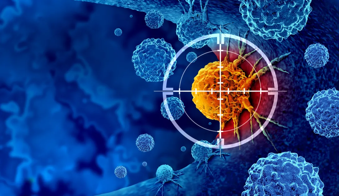
Illustration of cancer cells, with a highlighted tumor cell in the center targeted by a digital crosshair.

Advancing the frontiers of regenerative medicine means more than pushing scientific boundaries — it means improving and extending human life. The Regenerative Engineering and Medicine Center (REM) is a partnership with Georgia Tech, Emory University, and the University of Georgia (UGA) that supports this mission through inter-institutional collaborations in research in regenerative medicine.  

Since 2010, competitive peer-reviewed seed grants have been awarded annually to interdisciplinary teams with representation from at least two of the three institutions, leading to clinical trials, licensed technologies, start-up companies, and external funding for additional research. The Parker H. Petit Institute for Bioengineering and Bioscience (IBB) is excited to announce the 2026 REM Collaborative Seed Grant awardees: Melissa Kemp (Georgia Tech) and Rabindra Tirouvanziam (Emory); Yang Liu (UGA) and Yong Teng (Emory); and Steven Stice (UGA) and Zhexing Wen (Emory). 

Kemp and Tirouvanziam were awarded funding for their proposal, “Predicting Personalized Extracellular Vesicle (EV) Responses for Directed Myeloid‑Targeted Immunotherapy.” Their project combines computer modeling and lab‑grown lung tissue to better understand how immune cells communicate during lung infections and inflammation in different people. This research could help scientists design more precise, patient‑specific therapies for respiratory diseases, potentially improving treatments for conditions ranging from viral infections to chronic inflammation. 

"We are grateful for the support from REM that allows us to extend our labs into new, interdisciplinary research,” Kemp said. “This pilot project will allow us to develop and experimentally validate multicellular models of the lung environment. Our goal is to use our platforms to test potential therapeutics that operate by controlling communication between cell types." 

“It is wonderful to be supported by REM for this collaboration between Georgia Tech and Emory labs,” Tirouvanziam agreed. “We hope to turn this pilot into a large extramural project with a focus on novel immunotherapy.” 

Liu and Teng were awarded funding for their proposal, “AI‑Guided Profiling of Migratory Cancer Stem Cell Communication in Head and Neck Cancer.”  Their project aims to uncover how the most aggressive cancer stem cells move and “talk” to nearby immune and tissue cells, using advanced microfluidic tools and artificial intelligence to study how these cells help cancer spread and resist treatment.  Understanding these hidden communication pathways could lead to earlier detection of dangerous cancer cell types and inspire new therapies that prevent recurrence and improve survival for patients with head and neck cancer. 

“We combine microfluidic tools with artificial intelligence to monitor individual cancer cells in action and study how they interact with the immune microenvironment — capturing behaviors that are missed in bulk experiments and shedding light on how aggressive cancer cells escape therapy,” Liu said of the project.  

Stice and Wen were awarded funding for their application, “Use of Alzheimer’s Disease Organoids to Assess Mesenchymal Stromal Cell–Derived Extracellular Vesicles Mechanism of Action.”  Their project uses lab‑grown human brain organoids to study how tiny therapeutic particles called extracellular vesicles that are released by stem cells might reduce brain inflammation and protect neurons affected by Alzheimer’s disease.  Revealing how these vesicles work at a molecular level could help advance new treatments that go beyond symptom management and move toward slowing or preventing Alzheimer’s progression. 

“Extracellular vesicles (EVs) are used in the body to communicate with cells around an injury and are known to repair brain tissue in Alzheimer’s animal models,” Stice said.  “Understanding the signaling mechanisms used by EVs in Alzheimer’s brain organoids will directly lead to better EV manufacturing processes and potency for neurodegenerative diseases, and ultimately better therapies.” 

This year’s funded work illustrates how collaboration across institutions accelerates discoveries. Together, these teams are pushing healing technologies closer to real‑world impact, where they can make a tangible difference for patients affected by serious illness. 

News Contact

News Contact

Ashlie Bowman | Communications Program Manager

Parker H. Petit Institute for Bioengineering and Bioscience

Feb. 02, 2026
Hannah Youngblood
Raquel Lieberman

An estimated 4 million Americans have glaucoma, a group of eye diseases that can lead to irreversible blindness. Now, Georgia Tech is home to a Glaucoma Research Fund that will support cutting-edge work to understand and advance treatments for the disease.

The new initiative was sparked by ongoing research at Georgia Tech — and a Yellow Jacket connection: when Postdoctoral Research Fellow Hannah Youngblood’s work on exfoliation glaucoma (XFG) was featured by the BrightFocus Foundation, it caught the attention of Jennifer Rucker, an Alabama resident who was diagnosed with XFG several years ago.

Excited that the research could change outcomes for people like her — and proud that it’s happening at her husband Philip Rucker’s, EE 72, alma mater — Jennifer Rucker reached out to Youngblood and her advisor, School of Chemistry and Biochemistry Professor and Kelly Sepcic Pfeil, Ph.D. Chair Raquel Lieberman

“As the wife of a Georgia Tech graduate and an individual with pseudoexfoliation glaucoma, I was inspired to support the scientists whose efforts may help me and others,” Jennifer Rucker says. What followed was a meaningful dialogue and a shared sense of purpose — and the creation of the Georgia Tech Glaucoma Research Fund (Wreck Glaucoma! Fund). 

“It meant so much that Jennifer took the initiative to reach out to learn more about our research,” says Lieberman. “Moments like this remind me how deeply meaningful it is to connect with people in the broader community who are navigating glaucoma. Opportunities for such personal connections are rare, but they inspire and further motivate us to achieve our lab’s mission to improve the lives of individuals suffering from blindness diseases.”

A Personal Connection

Youngblood’s interest in glaucoma research also stems from a personal connection: her father was diagnosed with glaucoma as a young adult. Now, Youngblood studies the genetic and molecular factors behind XFG in the Lieberman research lab

“XFG is an aggressive form of the disease with no known cure,” Youngblood says. While scientists know that XFG is the result of abnormal accumulation of proteins in the eye, current treatments only address symptoms rather than treating the root cause of the disease.

“We know XFG is driven by protein buildup, but we still don’t know why it happens,” she explains. “My work studying specific genetic variants aims to uncover this.” 

The Genetics of Glaucoma

In particular, Youngblood is researching the role of LOXL1, a protein that plays a role in soft tissue throughout the body, including the eyes.

“Research has shown that people with variants in the genes responsible for this protein are more likely to have XFG,” she says. “That made me curious to see if the variants might be impacting the structure of the LOXL1 protein itself and how those variants might lead to disease.”

Youngblood is currently testing her theory in the lab. “My hope is that new insight into proteins like LOXL1 will bring us closer to treatments that address XFG at its source,” she says. “The new Georgia Tech Glaucoma Research Fund is a tremendous step forward in making that hope a reality.”

Support the Georgia Tech Glaucoma Research Fund

Please visit the Glaucoma Research Fund support page to give to this specific program. To discuss additional philanthropic opportunities, please contact the College of Sciences Development Team: development@cos.gatech.edu

Your investment ensures that these scholars and researchers have world-class resources, facilities, and mentors to excel in this critical work. Thank you for helping us shape the future.

News Contact

News Contact
Jan. 08, 2026
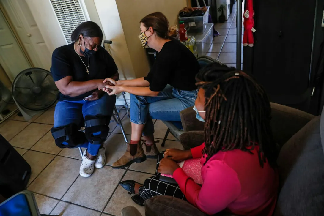
Lack of access to safe and affordable housing is harmful to health. Robert Gauthier/Los Angeles Times via Getty Images

Lack of access to safe and affordable housing is harmful to health. Robert Gauthier/Los Angeles Times via Getty Images

Health and medicine is more than just biological – societal forces can get under your skin and cause illness. Medical sociologists like me study these forces by treating society itself as our laboratory. Health and illness are our experiments in uncovering meaning, power and inequality, and how it affects all parts of a person’s life.

For example, why do low-income communities continue to have higher death rates, despite improved social and environmental conditions across society? Foundational research in medical sociology reveals that access to resources like money, knowledge, power and social networks strongly affects a person’s health. Medical sociologists have shown that social class is linked to numerous diseases and mortality, including risk factors that influence health and longevity. These include smoking, overweight and obesity, stress, social isolation, access to health care and living in disadvantaged neighborhoods.

Moreover, social class alone cannot explain such health inequalities. My own research examines how inequalities related to social class, race and gender affect access to autism services, particularly among single Black mothers who rely on public insurance. This work helps explain delays in autism diagnosis among Black children, who often wait three years after initial parent concerns before they are formally diagnosed. White children with private insurance typically wait from 9 to 22 months depending on age of diagnosis. This is just one of numerous examples of inequalities that are entrenched in and deepened by medical and educational systems.

Medical sociologists like me investigate how all of these factors interact to affect a person’s health. This social model of illness sees sickness as shaped by social, cultural, political and economic factors. We examine both individual experiences and societal influences to help address the health issues affecting vulnerable populations through large-scale reforms.

By studying the way social forces shape health inequalities, medical sociology helps address how health and illness extend beyond the body and into every aspect of people’s lives.

Protesters standing in front of a federal building, holding signs in the shape of graves reading '16 MILLION LIVES' and 'R.I.P. DEATH BY A THOUSAND CUTS,' wearing shirts that read 'MEDICAID SAVES LIVES'

Access to health insurance is a political issue that directly affects patients. Here, care workers gathered in June 2025 to protest Medicaid cuts. Tasos Katopodis/Getty Images for SEIU

Origins of Medical Sociology in the US

Medical sociology formally began in the U.S after World War II, when the National Institutes of Health started investing in joint medical and sociological research projects. Hospitals began hiring sociologists to address questions like how to improve patient compliance, doctor-patient interactions and medical treatments.

However, the focus of this early work was on issues specific to medicine, such as quality improvement or barriers to medication adherence. The goal was to study problems that could be directly applied in medical settings rather than challenging medical authority or existing inequalities. During that period, sociologists viewed illness mostly as a deviation from normal functioning leading to impairments that require treatment.

For example, the concept of the sick role – developed by medical sociologist Talcott Parsons in the 1950s – saw illness as a form of deviance from social roles and expectations. Under this idea, patients were solely responsible for seeking out medical care in order to return to normal functioning in society.

In the 1960s, sociologists began critiquing medical diagnoses and institutions. Researchers criticized the idea of the sick role because it assumed illnesses were temporary and did not account for chronic conditions or disability, which can last for long periods of time and do not necessarily allow people to deviate from their life obligations. The sick role assumed that all people have access to medical care, and it did not take into account how social characteristics like race, class, gender and age can influence a person’s experience of illness.

Patient wearing surgical mask sitting in chair of exam room, talking to a doctor

Early models of illness in medical sociology discounted the experience of the patient. Paul Bersebach/MediaNews Group/Orange County Register via Getty Images

Parsons’ sick role concept also emphasized the expertise of the physician rather than the patient’s experience of illness. For example, sociologist Erving Goffman showed that the way care is structured in asylums shaped how patients are treated. He also examined how the experience of stigma is an interactive process that develops in response to social norms. This work influenced how researchers understood chronic illness and disability and laid the groundwork for later debates on what counts as pathological or normal.

In the 1970s, some researchers began to question the model of medicine as an institution of social control. They critiqued how medicine’s jurisdiction expanded over many societal problems – such as old age and death – which were defined and treated as medical problems. Researchers were critical of the tendency to medicalize and apply labels like “healthy” and “ill” to increasing parts of human existence. This shift emphasized how a medical diagnosis can carry political weight and how medical authority can affect social inclusion or exclusion.

The critical perspective aligns with critiques from disability studies. Unlike medical sociology, which emerged through the medical model of disease, disability studies emerged from disability rights activism and scholarship. Rather than viewing disability as pathological, this field sees disability as a variation of the human condition rooted in social barriers and exclusionary environments. Instead of seeking cures, researchers focus on increasing accessibility, human rights and autonomy for disabled people.

A contemporary figure in this field was Alice Wong, a disability rights activist and medical sociologist who died in November 2025. Her work amplified disabled voices and helped shaped how the public understood disability justice and access to technology.

Structural Forces Shape Health and Illness

By focusing on social and structural influences on health, medical sociology has contributed significantly to programs addressing issues like segregation, discrimination, poverty, unemployment and underfunded schools.

For example, sociological research on racial health disparities invite neighborhood interventions that can help improve overall quality of life by increasing the availability of affordable nutritious foods in underserved neighborhoods or initiatives that prioritize equal access to education. At the societal level, large-scale social policies such as guaranteed minimum incomes or universal health care can dramatically reduce health inequalities.

People carrying boxes of food under a tent

Access to nutritious food is critical to health. K.C. Alfred / The San Diego Union-Tribune via Getty Images

Medical sociology has also expanded the understanding of how health care policies affect health, helping ensure that policy changes take into account the broader social context. For example, a key area of medical sociological research is the rising cost of and limited access to health care. This body of work focuses on the complex social and organizational factors of delivering health services. It highlights the need for more state and federal regulatory control as well as investment in groups and communities that need care the most.

Modern medical sociology ultimately considers all societal issues to be health issues. Improving people’s health and well-being requires improving education, employment, housing, transportation and other social, economic and political policies.The Conversation


This article is republished from The Conversation under a Creative Commons license. Read the original article.
News Contact

News Contact
Author:

Jennifer Singh, Associate Professor of Sociology, Georgia Institute of Technology

Media Contact:

Shelley Wunder-Smith
shelley.wunder-smith@research.gatech.edu

Dec. 16, 2025
Affectionally called "DragonCon for neuroscience," the annual Society for Neuroscience meeting is one of the largest academic conferences in the world.

Affectionally called "DragonCon for neuroscience," the annual Society for Neuroscience meeting is one of the largest academic conferences in the world.

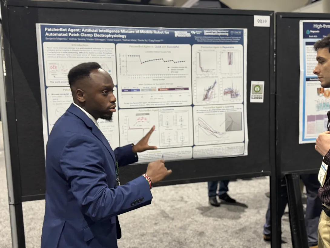
Benjamin Magondu, a graduate student in biomedical engineering, presented at SfN for the first time this year.

Benjamin Magondu, a graduate student in biomedical engineering, presented at SfN for the first time this year.

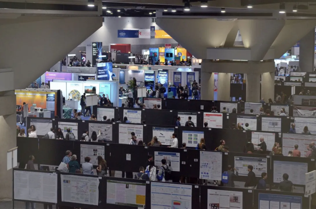
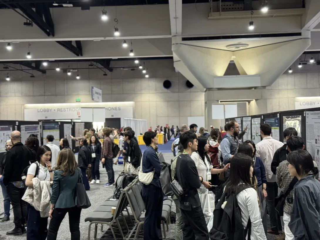
With hundreds of presentations happening simultaneously, the poster floor can be overwhelming at SfN — but for many, that's part of the draw.

With hundreds of presentations happening simultaneously, the poster floor can be overwhelming at SfN — but for many, that's part of the draw.

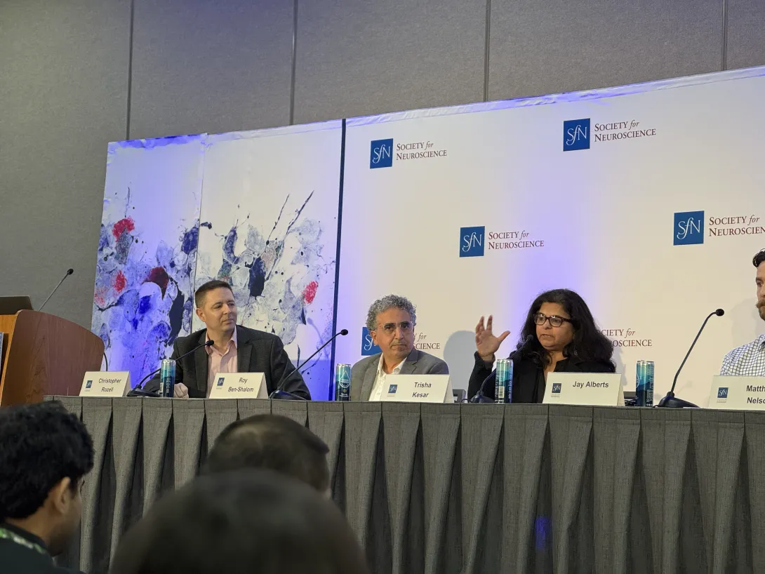
Trisha Kesar answers a question during the SfN press conference on AI in neuroscience, moderated by Chris Rozell.

Trisha Kesar answers a question during the SfN press conference on AI in neuroscience, moderated by Chris Rozell.

Imagine stepping into a space the size of multiple football fields — only instead of turf and goalposts, it’s filled with science. Every inch is alive with posters, equipment demos, and researchers sharing the latest breakthroughs.  

Welcome to the Society for Neuroscience (SfN) Conference, one of the largest scientific gatherings in the world, drawing more than 30,000 attendees to San Diego in November. According to Annabelle Singer, it is the place to be for neuroscientists. “If you want to know what is going on now in neuroscience, it is being talked about at SfN.” 

Singer is a McCamish Foundation Early Career Professor in the Wallace H. Coulter Department of Biomedical Engineering (BME) at Georgia Tech and Emory University. A frequent SfN attendee, she describes the meeting as “Dragon Con for neuroscience, with thousands of talks and posters going on simultaneously.” 

This year, Georgia Tech didn’t just show up — it made a statement with more than 60 presentations, a major outreach award, and a spotlight press conference. 

“Seeing Georgia Tech and INNS represented so strongly at SfN is exciting,” says Chris Rozell, executive director of Tech’s Institute for Neuroscience, Neurotechnology, and Society (INNS). “It reflects the incredible breadth of neuroscience and neurotechnology research happening across our campus and how our work is shaping conversations at the highest level.” 

Inside ‘Neuroscience Dragon Con’ 

Many conferences center around structured lectures, but at SfN, posters are the heart. You might find a senior researcher presenting groundbreaking findings right next to a first-time attendee sharing early results. This diversity is what makes the experience so valuable, says Singer. “Trainees get to talk directly with the scientist doing the work to get their questions answered, from wondering about future implications to clarifying technical details.” 

The scale of SfN can feel overwhelming, but for many, that’s part of the excitement. “There are so many different posters from so many different fields. It’s a lot to absorb, but it’s all very interesting,” said Benjamin Magondu, a biomedical engineering Ph.D. student presenting for the first time. “I’ve definitely learned at least 47 things by just walking 10 feet.” 

For students like Magondu, the experience is critical, says Biological Sciences Assistant Professor Farzaneh Najafi. “SfN has such a big scope, all the way from molecular to cognitive and computational systems. Especially for those deciding which direction of neuroscience they want to go into, it’s invaluable.” 

That breadth also fosters connections across disciplines. “Conferences are usually pretty niche,” noted Tina Franklin, a research scientist in BME. “You have your own field that you’re really good at, but it’s difficult to venture out and find new people who can help you figure out what comes next. This conference brings people from all different fields together with the common interest of neuroscience and brain research.” 

Leading the Charge 

Georgia Tech’s impact went beyond the conference floor. Ming-fai Fong, an assistant professor in BME, received the prestigious Next Generation Award, one of SfN’s education and outreach awards. The honor recognizes members who make outstanding contributions to public communication and education about neuroscience.  

“I’m certainly very grateful to the Society for Neuroscience for recognizing these types of contributions,” says Fong, who was recognized for her work supporting blind and visually impaired youth in Atlanta. “Rewarding outreach efforts reinforces my core belief that scientists and engineers can make an immediate impact on communities we care about through outreach. It’s a great parallel avenue to making a positive impact through research.” 

Building on this recognition, Georgia Tech was in the spotlight during one of SfN’s selective press conferences — a session on artificial intelligence in neuroscience moderated by Rozell, who is also the Julian T. Hightower Chair in the School of Electrical and Computer Engineering

During the SfN press event, Trisha Kesar, an associate professor in BME and adjunct faculty in the School of Biological Sciences, presented her research using AI to improve gait rehabilitation. Her work was among just 40 abstracts selected from more than 10,000 submissions for this honor, and one of five abstracts selected for the AI in neuroscience press conference. The project is a collaboration with Hyeok Kwon, a Georgia Tech computer science alumnus and an assistant professor in BME. 

“It’s exciting to see Georgia Tech and Atlanta emerging as hubs for neuroscience innovation,” said Kesar. “Being part of a press conference on AI in neuroscience shows how much our community is contributing to the future of brain research, and how collaboration across institutions can accelerate progress.” 

News Contact

News Contact

Writer and media contact:
Audra Davidson
Research Communications Manager
Institute for Neuroscience, Neurotechnology, and Society (INNS)

Presenter Dashboard:
Created by Joshua Preston, Communications Manager, College of Computing
Data collection by Audra Davidson, Hunter Ashcraft

Dec. 16, 2025
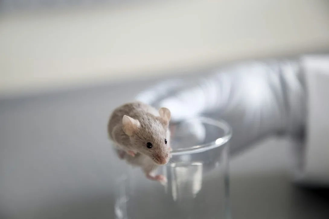
 Mice have complex visual systems that can clarify how vision works in people. Westend61/Getty Images

Mice have complex visual systems that can clarify how vision works in people. Westend61/Getty Images

Despite the nursery rhyme about three blind mice, mouse eyesight is surprisingly sensitive. Studying how mice see has helped researchers discover unprecedented details about how individual brain cells communicate and work together to create a mental picture of the visual world.

I am a neuroscientist who studies how brain cells drive visual perception and how these processes can fail in conditions such as autism. My lab “listens” to the electrical activity of neurons in the outermost part of the brain called the cerebral cortex, a large portion of which processes visual information. Injuries to the visual cortex can lead to blindness and other visual deficits, even when the eyes themselves are unhurt.

Understanding the activity of individual neurons – and how they work together while the brain is actively using and processing information – is a long-standing goal of neuroscience. Researchers have moved much closer to achieving this goal thanks to new technologies aimed at the mouse visual system. And these findings will help scientists better see how the visual systems of people work.

The Mind in the Blink of an Eye

Researchers long thought that vision in mice appeared sluggish with low clarity. But it turns out visual cortex neurons in mice – just like those in humans, monkeys, cats and ferrets – require specific visual features to trigger activity and are particularly selective in alert and awake conditions.

My colleagues and I and others have found that mice are especially sensitive to visual stimuli directly in front of them. This is surprising, because mouse eyes face outward rather than forward. Forward-facing eyes, like those of cats and primates, naturally have a larger area of focus straight ahead compared to outward-facing eyes.

Microscopy image of stacks of neurons

This image shows neurons in the mouse retina: cone photoreceptors (red), bipolar neurons (magenta), and a subtype of bipolar neuron (green). Brian Liu and Melanie Samuel/Baylor College of Medicine/NIH via Flickr

This finding suggests that the specialization of the visual system to highlight the frontal visual field appears to be shared between mice and humans. For mice, a visual focus on what’s straight ahead may help them be more responsive to shadows or edges in front of them, helping them avoid looming predators or better hunt and capture insects for food.

Importantly, the center of view is most affected in aging and many visual diseases in people. Since mice also rely heavily on this part of the visual field, they may be particularly useful models to study and treat visual impairment.

A Thousand Voices Drive Complicated Choices

Advances in technology have greatly accelerated scientific understanding of vision and the brain. Researchers can now routinely record the activity of thousands of neurons at the same time and pair this data with real-time video of a mouse’s face, pupil and body movements. This method can show how behavior interacts with brain activity.

It’s like spending years listening to a grainy recording of a symphony with one featured soloist, but now you have a pristine recording where you can hear every single musician with a note-by-note readout of every single finger movement.

Using these improved methods, researchers like me are studying how specific types of neurons work together during complex visual behaviors. This involves analyzing how factors such as movement, alertness and the environment influence visual activity in the brain.

For example, my lab and I found that the speed of visual signaling is highly sensitive to what actions are possible in the physical environment. If a mouse rests on a disc that permits running, visual signals travel to the cortex faster than if the mouse views the same images while resting in a stationary tube – even when the mouse is totally still in both conditions.

In order to connect electrical activity to visual perception, researchers also have to ask a mouse what it thinks it sees. How have we done this?

The last decade has seen researchers debunking long-standing myths about mouse learning and behavior. Like other rodents, mice are also surprisingly clever and can learn how to “tell” researchers about the visual events they perceive through their behavior.

For example, mice can learn to release a lever to indicate they have detected that a pattern has brightened or tilted. They can rotate a Lego wheel left or right to move a visual stimulus to the center of a screen like a video game, and they can stop running on a wheel and lick a water spout when they detect the visual scene has suddenly changed.

Mouse drinking from a metal water spout

Mice can be trained to drink water as a way to ‘tell’ researchers they see something. felixmizioznikov/iStock via Getty Images Plus

Mice can also use visual cues to focus their visual processing to specific parts of the visual field. As a result, they can more quickly and accurately respond to visual stimuli that appear in those regions. For example, my team and I found that a faint visual image in the peripheral visual field is difficult for mice to detect. But once they do notice it – and tell us by licking a water spout – their subsequent responses are faster and more accurate.

These improvements come at a cost: If the image unexpectedly appears in a different location, the mice are slower and less likely to respond to it. These findings resemble those found in studies on spatial attention in people.

My lab has also found that particular types of inhibitory neurons – brain cells that prevent activity from spreading – strongly control the strength of visual signals. When we activated certain inhibitory neurons in the visual cortex of mice, we could effectively “erase” their perception of an image.

These kinds of experiments are also revealing that the boundaries between perception and action in the brain are much less separate than once thought. This means that visual neurons will respond differently to the same image in ways that depend on behavioral circumstances – for example, visual responses differ if the image will be successfully detected, if it appears while the mouse is moving, or if it appears when the mouse is thirsty or hydrated.

Understanding how different factors shape how cortical neurons rapidly respond to visual images will require advances in computational tools that can separate the contribution of these behavioral signals from the visual ones. Researchers also need technologies that can isolate how specific types of brain cells carry and communicate these signals.

Data Clouds Encircling the Globe

This surge of research on the mouse visual system has led to a significant increase in the amount of data that scientists can not only gather in a single experiment but also publicly share among each other.

Major national and international research centers focused on unraveling the circuitry of the mouse visual system have been leading the charge in ushering in new optical, electrical and biological tools to measure large numbers of visual neurons in action. Moreover, they make all the data publicly available, inspiring similar efforts around the globe. This collaboration accelerates the ability of researchers to analyze data, replicate findings and make new discoveries.

Technological advances in data collection and sharing can make the culture of scientific discovery more efficient and transparent – a major data informatics goal of neuroscience in the years ahead.

If the past 10 years are anything to go by, I believe such discoveries are just the tip of the iceberg, and the mighty and not-so-blind mouse will play a leading role in the continuing quest to understand the mysteries of the human brain.The Conversation


This article is republished from The Conversation under a Creative Commons license. Read the original article.
News Contact

News Contact
Author:

Bilal Haider, Associate Professor of Biomedical Engineering, Georgia Institute of Technology

Media Contact:

Shelley Wunder-Smith
shelley.wunder-smith@research.gatech.edu

Dec. 11, 2025
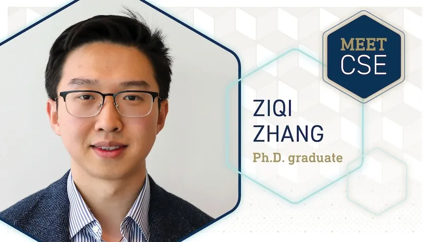
Meet CSE Ziqi Zhang

Ph.D. student Ziqi Zhang has built a career blending machine learning with single-cell biology. His work helps scientists study cellular mechanisms that advance disease research and drug development.

Though decorated with awards and appearances in leading journals, Zhang will achieve his greatest accomplishment tonight at McCamish Pavilion. He will join the Class of 2025 in walking across the stage, receiving diplomas, and graduating from Georgia Tech.

Before he “gets out” of Georgia Tech, we interviewed Zhang to learn more about his Ph.D. journey and where his degree will take him next. 

Graduate: Ziqi Zhang

Research Interests: Machine learning, foundational models, cellular mechanisms, single-cell gene sequencing, gene regulatory networks

Education: Ph.D. in Computational Science and Engineering

Faculty Advisor: School of CSE J.Z. Liang Early-Career Associate Professor Xiuwei Zhang

What persuaded you to study at Georgia Tech? 

I chose Georgia Tech because it is one of the top engineering institutions in the United States, known for its strength in machine learning and data science. The university offers exceptional research resources and the opportunity to work with leading scholars in my field. Georgia Tech also has very good research infrastructure. The Coda Building is one of the most well-designed and productive research environments I have experienced. Having access to such a space has been a genuine privilege.

How has working on your CSE degree helped you so far in your career?

Working toward my CSE degree has been instrumental in my career development. As an interdisciplinary program, CSE has equipped me with strong computational skills while also deepening my understanding of key application domains. This breadth of training has opened more opportunities during my job and internship searches. In addition, CSE community events, such as HotCSE, the weekly coffee hour, and faculty recruiting activities, have helped me strengthen my scientific communication skills, which are essential for my long-term career growth.

What research project from Georgia Tech are you most proud of?

My favorite research project was scMoMaT, a matrix tri-factorization algorithm for single-cell data integration. I invested a significant amount of time and effort into this work, iterating on the model many times. I’m very proud that it ultimately evolved into a clean, robust, and elegant algorithm.

What advice would you give someone interested in graduate school?

It is important to find an advisor who is supportive and genuinely invested in your career development. A Ph.D. is not an easy journey, and you will inevitably encounter challenges along the way. Having an advisor who can provide thoughtful guidance and dedicated mentorship is one of the most crucial factors in helping you navigate those difficulties.

What is your most favorite memory from Georgia Tech?

CSE’s new student campus visit day every year was one of my favorite times of the year. It was always fun to meet new people, have good food, and enjoy the beautiful view from the Coda rooftop.

What are your plans after graduation?

I plan to keep working in academia after graduation. I’m on the job hunt, currently applying for positions and preparing for interviews.

News Contact

News Contact

Bryant Wine, Communications Officer
bryant.wine@cc.gatech.edu

Dec. 10, 2025
Yunan Luo NSF CAREER Award

Proteins, including antibodies, hemoglobin, and insulin, power nearly every vital aspect of life. Breakthroughs in protein research are producing vaccines, resilient crops, bioenergy sources, and other innovative technologies.

Despite their importance, most of what scientists know about proteins only comes from a small sample size. This stands in the way of fully understanding how most proteins work and unlocking their full potential.

Georgia Tech’s Yunan Luo believes artificial intelligence (AI) could fill this knowledge gap. The National Science Foundation agrees. Luo is the recipient of an NSF Faculty Early Career Development (CAREER) award. 

“So much of biology depends on knowing what proteins do, but decades of research have concentrated on a relatively small set of well-studied proteins. This imbalance in scientific attention leads to a distorted view of the biological landscape that quietly shapes our data and our algorithms,” Luo said.

“My group’s goal is to build machine learning (ML) models that actively close this gap by generating trustworthy function predictions for the many proteins that remain understudied.”

[Related: Yunan Luo to use AI for Protein Design and Discovery with Support of $1.8 Million NIH Grant]

In his proposal to NSF, Luo coined this rich-get-richer effect “annotation inequality.” 

One problem of annotation inequality is that it slows progress in disease prognosis, drug discovery, and other critical biomedical areas. It is challenging to innovate the few proteins that scientists already know so much about. 

A cascading effect of annotation inequality is that it diminishes the effectiveness of studying proteins with AI.  

AI methods learn from existing experimental data. Datasets skewed toward well-known proteins propagate and become entrenched in models. Over time, this makes it harder for computers to research understudied proteins. 

“Protein annotation inequality creates an effect analogous to a vast library where 95% of patrons only read the top 5% popular books, leaving the rest of the collection to gather dust,” Luo said.

“This has resulted in knowledge disparities across proteins in current literature and databases, biasing our understanding of protein functions.”

The NSF CAREER award will fund Luo with over $770,000 for the next five years to tackle head-on the problem of protein annotation inequality.

Luo will use the grant to build an accurate, unbiased protein function prediction framework at scale. His project aims to:

  • Reveal how annotation inequality affects protein function prediction systems
  • Create ML techniques suited for biological data, which is often noisy, incomplete, and imbalanced  
  • Integrate data and ML models into a scalable framework to accelerate discoveries involving understudied proteins

More enduring than the ML framework, Luo will leverage the NSF award to support educational and outreach programs. His goal is to groom the next generation of researchers to study other challenges in computational biology, not just the annotation inequality problem.

Luo teaches graduate and undergraduate courses focused on computational biology and ML. Problems and methods developed through the CAREER project can be used as course material in his classes.

Luo also championed collaboration with Georgia Tech’s Center for Education Integrating Science, Mathematics, and Computing (CEISMC) in his proposal. 

Through this partnership, local high school teachers and students would gain access to his data and models. This promotes deeper learning of biology and data science through hands-on experience with real-world tools.  

Luo sees reaching students and the community as a way of paying forward the support he received from Georgia Tech colleagues. 

“I am incredibly grateful for this recognition from the NSF,” said Luo, an assistant professor in the School of Computational Science and Engineering (CSE). 

“This would not have been possible without my students and collaborators, whose hard work laid the groundwork for this proposal.”

Luo praised CSE faculty members B. Aditya Prakash, Xiuwei Zhang, and Chao Zhang for their guidance. All three study machine learning and computational bioscience, two of CSE’s five core research areas

Luo also thanked Haesun Park for her support and recommendation for the CAREER award. Park is a Regents’ Professor and the chair of the School of CSE.

News Contact

News Contact

Bryant Wine, Communications Officer
bryant.wine@cc.gatech.edu

Nov. 24, 2025
A tall white man wearing a blue GT-branded polo standing next to a slightly shorter man wearing a UGA-branded red polo. They're smiling and both holding a football.

Tim Lieuwen and Chris King

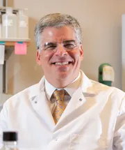
A man in a white lab coat and glasses, with a gold tie

Andrés J. García

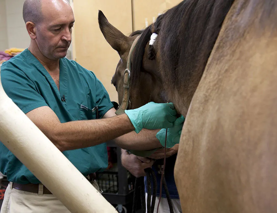
A man wearing teal surgical cloges and a green scrubs top, next to a light brown horse

John Peroni

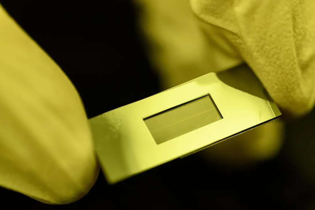
The Dynamic Mass Spectrometry Probe developed to monitor the health of living cell cultures (photo credit: Rob Felt)

The Dynamic Mass Spectrometry Probe developed to monitor the health of living cell cultures (photo credit: Rob Felt)

A smiling woman with long brown hair, wearing a black t-shirt and a floral cardigan

Sarah Farmer

If you’ve lived in Georgia long enough, you’ve almost certainly heard the friendly jabs tossed across divided Thanksgiving tables. On one side, a smirk and a mention of the “North Avenue Trade School.” On the other, a pointed retort: “To hell with Georgia.”

Few rivalries run deeper than the one known as “Clean, Old-Fashioned Hate,” the annual showdown between Georgia Tech and the University of Georgia (UGA). On Friday afternoon, November 28, the two will face off in one of the most anticipated matchups in years. These teams don’t like each other, and for a few hours every year, neither do friends, families, and even significant others.

Off the field, however, the schools are proving that collaboration, not competition, is the schools’ true strength.

For more than a century, Georgia’s flagship universities have united around complementary strengths, tackling the state’s biggest challenges together. That starts with making Georgians healthier.

“When Georgia Tech and UGA combine their strengths, together we create solutions that neither institution could achieve alone,” said Tim Lieuwen, executive vice president for Research at Georgia Tech. “These collaborations accelerate innovation in healthcare, improve lives across our state, and demonstrate that partnership — not rivalry — is Georgia’s most powerful tradition."

“The common denominator between these two great institutions is the populations they serve,” said Chris King, interim vice president for Research at UGA. “We have a duty to find solutions that help improve the quality of life for all Georgians, and that’s what these partnerships are all about.”

From programs like the Georgia Clinical and Translational Science Alliance (Georgia CTSA) to the National Science Foundation’s Engineering Research Center for Cell Manufacturing Technologies (CMaT), researchers at UGA and Georgia Tech are setting rivalries aside to build lasting partnerships that fuel innovation and expand the workforce to meet the state’s needs.

Pushing Cell Therapy Across the Goal Line
CMaT is an NSF-funded consortium of more than seven universities and 40 member companies. At Georgia Tech and UGA, teams are conducting many early stage translational projects to improve manufacturing of cell-based therapeutics.

One joint project between Andrés García, executive director of Georgia Tech’s Parker H. Petit Institute for Bioengineering & Bioscience, and John Peroni, the Dr. Steeve Giguere Memorial Professor in Large Animal Medicine in UGA’s College of Veterinary Medicine, addresses treatment of bacterial infections that can follow bone repair surgeries.

Bone fractures and non-union defects often require surgical implants, but 1-5% are compromised by bacterial infection, costing hospitals more than $1.9 billion annually. Current treatments are limited to sustained, high doses of antibiotics, which are less effective and can generate antibiotic-resistant bacteria. García and Peroni are engineering synthetic biomaterials that locally deliver antimicrobial agents to eliminate infections and promote bone repair.

Steven Stice, D.W. Brooks Distinguished Professor and Georgia Research Alliance Eminent Scholar at UGA’s Regenerative Bioscience Center, is also working with Georgia Tech’s Andrei Fedorov, professor and Rae S. and Frank H. Neely Chair in the George W. Woodruff School of Mechanical Engineering, to improve the quality and control of producing natural, cell-derived healing materials for regenerative medicine.

Adult cells secrete tiny, bubble-like vesicles that help other cells heal and regenerate tissue. Stice developed methods to boost vesicle production, while Fedorov created a probe that accelerates the process.

“Cells simply don’t secrete these healing vesicles in the quantities needed for scalable, clinical-grade treatments,” said Stice, UGA lead and co-principal investigator for CMaT. “Our collaborative work changes that, accelerating production in a way that finally makes large-scale regenerative therapies feasible.”

“Georgia Tech and UGA's collective commitment to advancing science and technology exceeds the intensity of our athletic rivalry,” Fedorov said. “Together, we’re advancing cell and therapy biomanufacturing to develop lifesaving treatments for the most devastating diseases.”
 
Georgia Tech’s Francisco Robles and UGA’s Lohitash Karumbaiah are using manufactured T cells to target cancer. Robles, who leads the Optical Imaging and Spectroscopy Lab in the Wallace H. Coulter Department of Biomedical Engineering, developed quantitative Oblique Back-illumination Microscopy (qOBM) to monitor tumor growth in real time. The method allows scientists to visualize patient-derived glioblastoma cell clusters generated in the Karumbaiah Lab, tracking tumor structure and behavior at various stages.

“Assessing therapeutic potency is often complex, costly, and ineffective for solid tumors,” Karumbaiah said. “qOBM simplifies the process by providing real-time, label-free monitoring of therapeutic efficacy against 3D solid tumors.”   

The work could help doctors personalize cancer treatments by providing early, detailed signs of whether a therapy is working.

“This technique is more compact and affordable and lets us watch T cells attack cell cultures in real time,” Robles said. “This breakthrough could transform how we study disease and screen new treatments.”

A Playbook for Local Healthcare
Created in 2007 by the National Institutes of Health, Georgia CTSA is one of several NIH-funded national partnerships advancing new health therapeutics and practices. Since 2017, it has comprised UGA, Georgia Tech, Emory, and the Morehouse School of Medicine. The alliance’s reach extends far beyond campus borders, bringing together researchers, clinicians, professional societies, and community and industry partners to identify local health challenges and translate research into practical solutions.

And out of this alliance have come many collaborative studies among CTSA’s members.

One, the Georgia Health Landscape Dashboard, is a tool to identify local health gaps and connect regional health professionals or policymakers with the researchers who can best address their community’s challenges. UGA College of Family and Consumer Sciences Associate Professors Alison Berg and Dee Warmath, along with community health engagement coordinator Courtney Still Brown, are working with Georgia Tech’s Jon Duke, director of the Center for Health Analytics and Informatics at the Georgia Tech Research Institute and a principal research scientist in the School of Interactive Computing.

The dashboard has already helped match researchers with communities by combining epidemiological data with “community voice” insights through surveys of residents and local leaders.

For example, when examining diabetes data, the dashboard indicates Randolph County has the state’s highest prevalence, despite declining by about 8% between 2021-24. Meanwhile, Treutlen County’s rate increased 29.2% during the same period. Perhaps Treutlen’s need for diabetic care is a growing concern, while Randolph’s is being addressed. And perhaps Hancock County, which ranks diabetes its top priority in the community voice category, is in search of immediate solutions.

“The Landscape Dashboard is a fantastic example of how the unique expertise found at Georgia Tech and UGA can be brought together to create something truly valuable for all Georgia,” Duke said. “By bringing together a range of data sources and health analytics approaches, this collaboration has created a tool that delivers novel insights into health, community, and policy across the state.”

Supported by UGA Cooperative Extension and the Biomedical and Translational Sciences Institute, the project leverages a network of agents in every county across the state. Warmath said the project’s strength lies in its ability to connect research with real-world needs.

“To build a community-responsive ecosystem for biomedical research, scientists must recognize local needs, share progress with communities to foster trust and acceptance, recruit clinicians and industry partners, and strengthen the relationships between patient and caregiver,” Warmath said.

Teaming Up for Maternal Health
Warmath and a team of researchers at UGA, Georgia Tech, and Emory are also collaborating on an NIH-funded project uniting experts in maternal health, biostatistics, and consumer science to explore how wearable technologies could improve delivery-room care.

During childbirth, clinicians monitor countless maternal and fetal vitals — contractions, heart rates, oxygen levels, kidney function, and more. What new insights, the researchers asked, could advanced wearable technologies offer in the delivery room, and what barriers might prevent their use?

Using nationwide surveys and focus groups, the team gathered information from a representative sample of pregnant, postpartum, and reproductive-age women, as well as healthcare professionals, to examine acceptance of wearable health technologies during labor and delivery. In their analysis of this rich data source, the team is identifying key variables that reveal gaps in technology acceptance and the unique needs of diverse maternal populations.

Each partner institution brings unique expertise. At Emory, principal investigator Suchitra Chandrasekaran contributes clinical insights from direct patient care. At UGA, Warmath applies her knowledge in consumer science to analyze end-user motivation, attitudes, and behaviors. At Georgia Tech, experts like Sarah Farmer in the Center for Advanced Communications Policy’s Home Lab facilitate large-scale data collection.

With data collection now complete, the team is analyzing results to inform future design and deployment of wearable technologies.
“Each school has a different perspective,” Farmer said. “It’s not as simple as one school does this but doesn’t do that. Each has their expertise, but they offer different perspectives and different resources that, when pooled, can make our research that much more effective.”

Whether advancing maternal health, mapping Georgia’s health needs, or engineering next-generation therapies, UGA and Georgia Tech continue to prove that collaboration is Georgia’s strongest tradition. Further, the undergraduate and graduate students who work in these labs and others represent the state’s highly skilled workforce of tomorrow.

“When our institutions work together, Georgia wins,” Warmath said.

By David Mitchell

News Contact

News Contact

For media inquiries:
Angela Bajaras Prendiville
Director of Media Relations
media@gatech.edu

Nov. 10, 2025
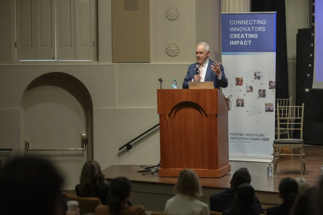
President Ángel Cabrera of Georgia Tech stands at a podium and delivers a speech.

Imagine a world where pediatric gastrointestinal disease could be diagnosed by swallowing a capsule-sized camera, where heart defects could be corrected by biodegradable implants, and where ADHD could be diagnosed through virtual reality. Georgia Tech and its partners are helping bring these world-changing ideas to life. 

On Nov. 5 – 6, Georgia Tech hosted the Pediatric Healthcare Innovation Summit 2025 (PHIS), a one-of-a-kind event that brought champions of children’s health together to share knowledge, facilitate collaborative initiatives, and accelerate medical innovation. The summit was co-presented by the Georgia Tech Pediatric Innovation Network (PIN), the International Society for Pediatric Innovation (ISPI), and the FDA-funded Pediatric Device Consortia (PDC).

The event included a tour of the new Arthur M. Blank Hospital, technology showcases, workshops, panel discussions, a poster session, and a pitch competition where companies were awarded funding from the Pediatric Device Consortia. 

“Georgia Tech is committed to advancing medicine, but in particular pediatric medicine, which is normally underfunded compared to adult healthcare,” Georgia Tech President Ángel Cabrera said. “We are committed to playing our part, and we're doing that in partnership with the best organizations, combining our engineering skills with clinical partners who understand the most important needs of children.”

Cabrera was a guest speaker for the event, which took place at two locations across campus: the newly opened Science Square and the Historic Academy of Medicine. He emphasized that championing causes such as pediatric healthcare innovation not only aligns with Georgia Tech’s mission, but also with the vision surrounding the new infrastructure being built across campus.

“We're committed to turning our city and our neighborhood into a hub of innovation, and the area of life sciences is one of those areas that we are supporting — including our new Science Square neighborhood, which is devoted to precisely this,” he said.

Though industry events happen every year, what makes PHIS unique is its goal of uniting not only clinicians and healthcare administrators, but also researchers, investors, and entrepreneurs.  Attendees are united around a shared goal of solving systemic problems and, ultimately, saving and improving the lives of children. Julia Kubanek, Georgia Tech’s Vice President for Interdisciplinary Research, said that this collaborative approach provides a unique opportunity to progress ideas and technologies that impact the industry.

“Particularly in the pediatric space, the market is relatively small. When you have a specialized pediatric technology, it's sometimes difficult to get the resources to advance that into clinical trials and into products that can go to market,” she said. “This environment that the summit creates is a supportive one for solving those problems and advancing life-saving research.”

While this was the third year that the event featured a pitch competition, it was the first year that winners were awarded monetary prizes. By bringing startups and investors together, the PHIS plays a vital role in getting impactful research from conceptual to consumer ready. This year’s winners included: Luminoah in first place, Rhaeos in second, and AcQumen Medical in third.

Though the event does encourage friendly competition, the ultimate goal remains to improve the lives of children and their families through collaboration, thought leadership, and innovation.

“Our north star is taking care of children,” Anthony Chang, founder of ISPI, said in his opening remarks. “I think we underestimate how much we learn together. I look at our jobs not as jobs but as a special calling — taking care of children.”

In addition to PIN, ISPI, and PDC, the event was sponsored by Georgia Tech’s Office of Corporate Engagement, Shriner’s Children’s Research Institute, Children’s Healthcare of Atlanta, the Georgia Department of Economic Development, the Georgia Research Alliance, and the International Children’s Advisory Network, among others.

News Contact

News Contact

Ashlie Bowman

Research Communications Program Manager

Subscribe to Biotechnology, Health, Bioengineering, Genetics